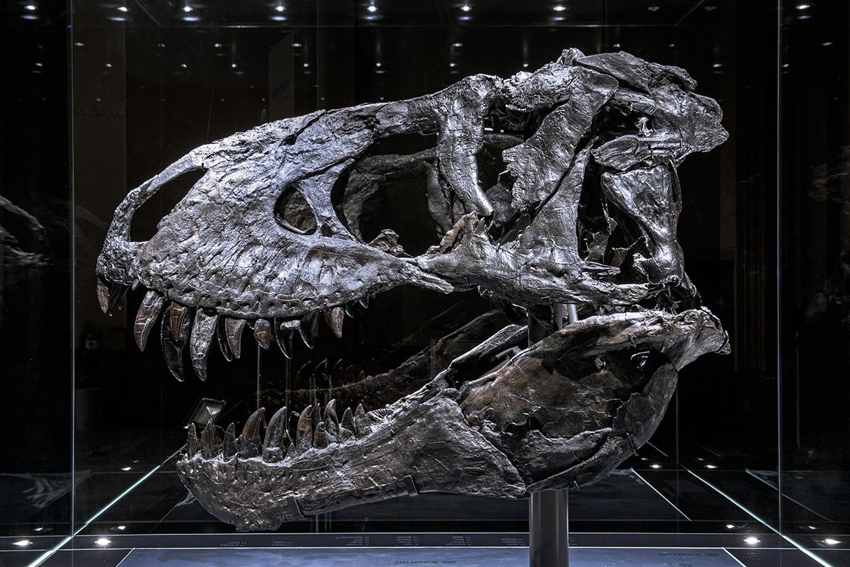
Scientists of the Museum für Naturkunde Berlin and physicians are facing new challenges and tested the workflow and image quality of clinical computed tomography in a study compared to photogrammetry on the skull of the Tyrannosaurus rex Tristan Otto. The imaging methods are suitable for pathological studies and for better recognition of clinical diagnoses. Tristan Otto will be on display at least until 28 February 2019, before the skeleton embarks on his journey to Copenhagen's Natural History Museum in spring. A precise departure date has not yet been set.
Imaging is crucial to gather scientific data in palaeontology. Photogrammetry is currently a frequently used technique for surface imaging, producing high-quality 3D surface data. Clinical computed tomography (CT) scanners are interesting for palaeontological research because of their high availability and the potential to image internal structures in addition to the surface.
This study with the participation of palaeontologists Daniela Schwarz and Oliver Hampe from the Museum für Naturkunde Berlin examines the technical effort, workflow and image quality of clinical computed tomography compared to photogrammetry. As an excellent example the skull of Tristan Otto, the Tyrannosaurus rex from the Upper Cretaceous of Montana/USA, of which 47 bone elements have been preserved, was used.
The study could be facilitated because of the good preservation of the individual skull bones. In contrast to many other Tyrannosaurus skulls, the skull bones of Tristan Otto were found isolated and later assembled into a skull. Thus all bones could be examined individually with both techniques – an examination of the completely assembled skull would not have been feasible because of size and weight. In the following, 3D models were created from both CT and photogrammetry data. Scanning times, data volumes, the overall radiation exposure, and the costs for the different procedures were compared. This study shows that a clinical CT scanner can be used for imaging even large palaeontological objects with high density. In comparison to CT scanning, the data-capture effort of photogrammetry is directly linked to the size and color of the specimen and to the complexity of its shape. While those factors influence the photogrammetry-based 3D model and the quality of its details, the CT scan is mostly free of these variables. Unlike the acquisition and calculation time in photogrammetry the CT scanning time for large and small objects measures roughly the same, as this method is independent of the specimen’s shape and complexity.
Since the spatial resolution in computed tomography is considerably lower than in photogrammetry, while the latter lacks the ability to reveal internal structures, neither technique can replace the other. On the contrary, CT scanning and photogrammetry complement each other and can be used not only in palaeontological research but also for comprehensive clinical imaging such as 3D simulation in reconstructive plastic surgery.
