In this laboratory complex of the Museum für Naturkunde Berlin, researchers investigate the chemical composition and structure of rocks, meteorites, minerals and fossils. The objects come from collections, expeditions and experiments. The material-related research is based on various imaging and chemical-physical methods. The laboratory complex is located on one floor of the museum’s main building for technical and organisational reasons. It features several, complementary large appliances. In many cases, answers to research questions can only be found in the micrometre range. In addition to the museum's working groups, the laboratory complex is also available to guests: Researchers from other institutions, young scientists and students from the near-by universities. Guided tours for visitors provide insights into the research.
Equipment
Scanning electron microscope JEOL JSM-6610LV with LaB6 cathode, Cl detector and a Bruker EBSD system CrystAlign 400
Head of laboratory: Dr. Lutz Hecht
Laboratory employee: Kirsten Born
The scanning electron microscope (SEM) is used for the visualisation of structures in the micrometre range and the chemical analysis of rocks, minerals and fossils. The large sample chamber holds objects up to ten centimetres in size. The SEM has a low vacuum mode so that even uncoated or not fully vacuum-resistant samples can be examined.
Electron microprobe with field emission cathode JEOL JXA-8500F, 5 WDX spectrometer, Bruker EDX System Quantax
Head of laboratory: Dr. Lutz Hecht
Laboratory employee: Dr. Felix Kaufmann
The electron microprobe is used mainly to investigate the chemical composition of rocks and mineral phases in the centimetre down to the micrometre range quantitatively. Typically, highly polished thick or thin slices are used for this purpose. Highly vacuum resistant fossil specimens can also be investigated. Equipped with a field emission cathode, the microprobe can image structures with particularly high resolution and can be used for chemical analyses as well as the recognition of elemental distribution patterns down to the nanometre range. Using an e-flash EDS detector, element distribution maps in thin slices of rock can be created and special phases can be found in a short time.
Micro X-Ray Fluorescence (µXRF) Lab
Head of laboratory: Dr. Christopher Hamann
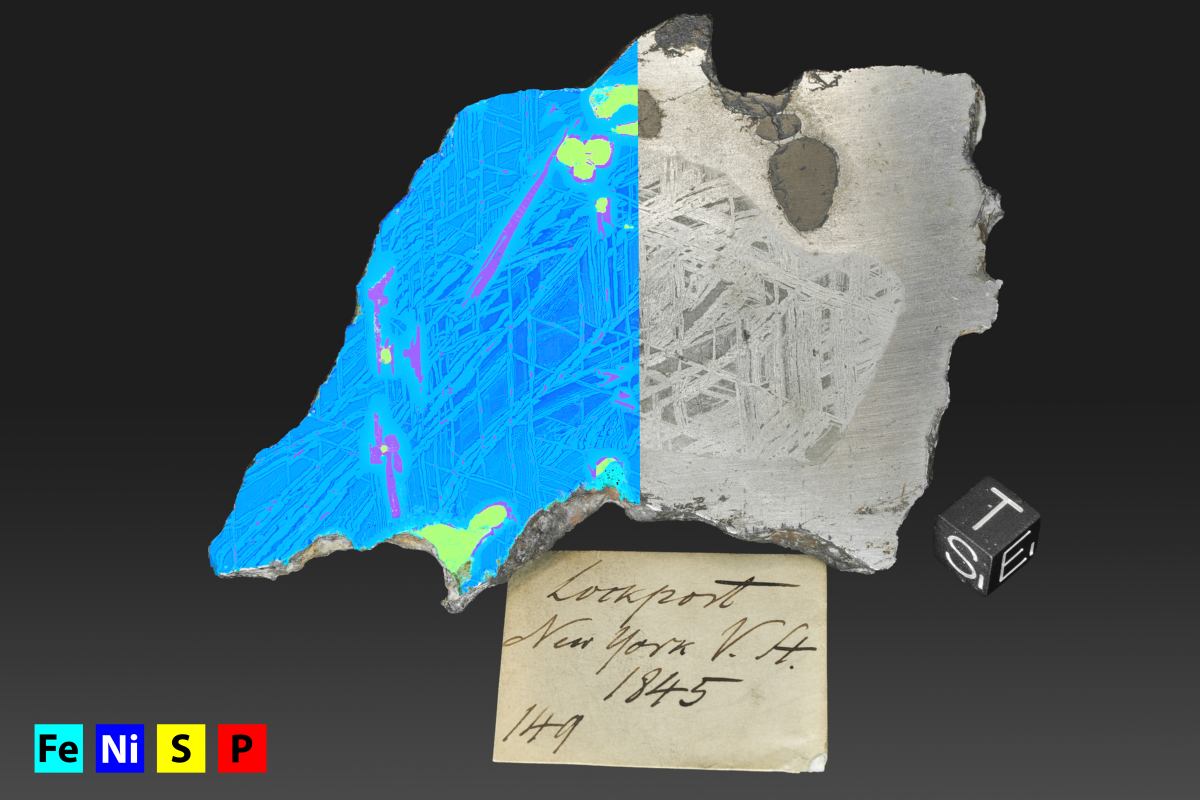
The Micro X-Ray Fluorescence (µXRF) Lab houses two spectrometers used for element distribution maps and quantitative analysis of the main and trace elements. The Bruker Tornado M4 plus can analyse samples up to 16x20 cm and runs under vacuum to allow detection of low mass elements. The lowest spot size is 20 µm. The Bruker Jetstream M6 can analyse large samples with a maximum mapping size of 60x80 cm. It usually runs under atmosphere (no detection of low mass elements). The spot size is variable (lowest 200 µm). In addition, the µXRF Lab houses a Bruker TRACER IV hand-held X-Ray fluorescence spectrometer that allows semiquantitative to quantitative (if calibrated against a known standard) analysis of centimetre-sized objects on-site in the field.
Raman spectrometer Horiba LabRam
Head of laboratory: Dr. Christopher Hamann
Laboratory employee: Kirsten Born
The Raman spectrometer is used to structurally characterize and identify mineral phases, glasses and organic substances. It is suitable for almost all areas of object-oriented research. Raman spectroscopy provides information on the molecular or crystalline structure of samples down to the micrometre range. Flexible fibre optics allow for non-destructive examinations of surfaces of larger collection objects. The Raman spectrometer is equipped with three lasers of different wavelength (an air-cooled solid-state laser of 473 nm wavelength, a HeNe laser of 633 nm wavelength, and an air-cooled laser diode of 785 nm wavelength), an Olympus optical microscope, and a motorized sample stage.
X-ray fluorescence spectrometer Bruker S8 Tiger
Laboratory Manager: Dr. Ralf-Thomas Schmitt
Laboratory employee: Kathrin Krahn
The X-ray sequence spectroscopy laboratory is used for analysis of the main and trace elements of rocks and minerals with element concentrations as low as the parts per million range. The analyser can be used on-site as well as for sample-based research projects for analyses and also for the pre-selection of samples for highly specific geochemical investigations (INNA, REE and PGE analysis). In the laboratory, rocks can be processed with jaw crushers and various rock mills.
Cathodoluminescence microscope
Head of laboratory: Dr. Lutz Hecht
Laboratory employee: Kirsten Born
The cathode ray of this microscope can make mineral phases glow. This effect is mainly used to investigate mineral generations and zoning in minerals and melts.
Mineralogical preparation
Head of laboratory: Dr. Ansgar Greshake
Laboratory employee: Karen Helm
Mineralogical preparation covers the spectrum of coarse and fine sawing work, polished sections and thin sections polished on one or both sides. Surface and thin sections can be produced in Giessen format or as one-inch cylindrical sections. The laboratory is equipped with various rock saws, an inner diameter saw, two GNM grinding machines, two Logitech polishing machines and a polishing machine for vibration polishing.
Photo: Cambria meteorite, with visible Widmanstätten pattern in the centre. The left half of the image shows the element distribution map for iron, nickel, sulphur and phosphorus, acquired using the in-house micro X-Ray fluorescence lab. H. Knöfler.
