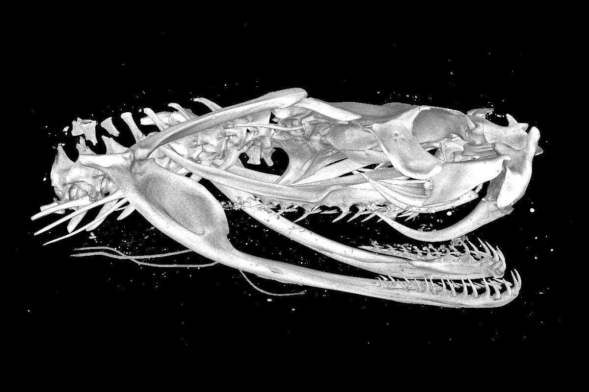
In the Micro-CT laboratory, biological, palaeontological and geological objects are analysed using computer-assisted methods. The laboratory is a hub both for research at the museum and for the digitisation of the scientific collections. In addition to various devices for three-dimensional scanning, the laboratory has 12 powerful workstations for processing and analysing the raw data using special 3D volume processing software. The laboratory is used by a number of different working groups and the research projects range from evolutionary studies to taxonomic and palaeontological studies to geological projects, e.g. in the context of impact research. Services for external users are also offered.
Methodology (selection):
- X-ray tomography
- 3D Photogrammetry
- 3D surface scan
Equipment (selection):
Phoenix Nanotom S - X-ray tomograph
Technical direction: Kristin Mahlow
For X-ray-based examination and three-dimensional recording of external and internal structures. The maximum size of the objects is 15 centimetres, resolutions down to 0.5 micrometres can be achieved. The 3D volume processing is usually done with the software “Volume Graphics Studio Max”, but also with “Amira”.
Artec Spider, Artec EVA - hand-held scanner
Technical direction: Kristin Mahlow
For three-dimensional scanning of external surface structures. The two Artec devices are portable and can be taken into collections in order to allow non-destructive examination of objects. A special software developed by Artec as well as a powerful laptop enable quick representations of structures, so that many objects can be captured three-dimensionally in a short time. The objects can be five centimetres to 1.5 metres (spider) or 20 centimetres to six metres in size (EVA).
Soft tissue staining laboratory
Technical direction: Kristin Mahlow
Preparation of biological samples for the virtual representation of soft tissues through X-ray analysis. Common staining protocols of DiceCT (iodine) or tungstic acid (PTA) staining for muscle or cartilage structure analysis.
Applications (examples):
- Using 3D X-ray tomography and the development of new techniques for the visual representation of soft tissue structures, it was possible to show for the first time how the trunk muscles of snake-like reptiles change evolutionarily when the limbs are lost.
Westphal N, Mahlow K, Head JJ, Müller J (2019) Pectoral myology of limb-reduced worm lizards (Squamata, Amphisbaenia) suggests decoupling of the musculoskeletal system during the evolution of body elongation. BMC Evolutionary Biology 19:16. https://doi.org/10.1186/s12862-018-1303-1
Gignac PM, Kley NJ, Clarke JA, Colbert MW, Morhardt AC, Cerio D, Cost IN, Cox PG, Daza J. D, Early CM, Echols MS, Henkelman RM, Herdina AN, Holliday CM, Li Z, Mahlow K, Merchant S, Müller J, Orsbon CP, Paluh DJ, Thies ML, Tsai HP, Witmer LM (2016), Diffusible iodine-based contrast-enhanced computed tomography (diceCT): an emerging tool for rapid, high-resolution, 3-D imaging of metazoan soft tissues. Journal of Anatomy, 228: 889-909. doi: 10.1111/joa.12449
- X-ray tomography has helped to identify a previously unknown family of frogs in the rainforests of West African Guinea. This discovery underlines the importance of Guinea as a biodiversity hotspot.
Barej MF, Schmitz A, Günther R, Loader SP, Mahlow K, Rödel MO (2014) The first endemic West African vertebrate family - a new anuran family highlighting the uniqueness of the Upper Guinean biodiversity hotspot. Frontiers in Zoology, 11, p. 8, 2014 doi:10.1186/1742-9994-11-8
- 3D surface scans of skulls of fossil and recent African antelopes are used to investigate how different ecological adaptations have affected the evolution and appearance of the different species and their ancestors.
