Topic and objectives
This permanent research field studies all kinds of pathologies that manifest in the skeleton of fossil vertebrates. Palaeopathology offers the deep time perspective of disease and asks question about the evolutionary origin and history of disease. What kinds of disease affected extinct vertebrates millions of years ago? Can we recognize the basically same types, or different ones than in today’s vertebrates including humans? How did diseases change with the evolution of organisms? How did bone healing and regenerative capacity evolve in the history of vertebrates? What inferences can we draw from ancient diseases and injuries concerning lifestyle and behavior of long extinct animals?
Methodology
We investigate pathologically altered fossil bone macroscopically and microscopically, by micro-CT, synchrotron X-ray phase contrast tomography, and histological thin sections.
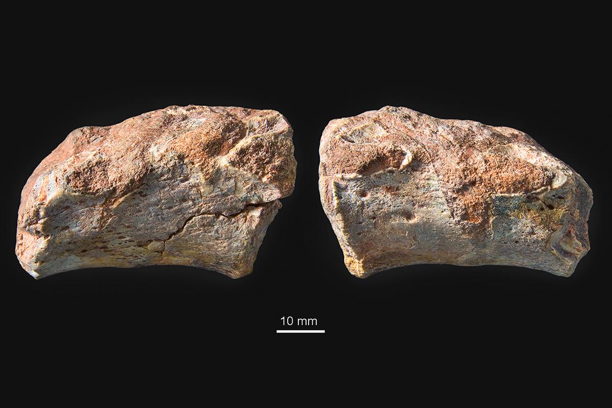
photograph on top: Two fused vertebral bodies of the Late Triassic temnospondyl amphibian Metoposaurus algarvensis with an intercalated hemivertebra on the left side. Top: specimen in left lateral view showing three articulation facets for the ribs. Bottom: specimen in right lateral view with two articulation facets. Hemivertebra is a congenital malformation in which a vertebra develops only on either the left or right side. This pathology is also known in human medicine.
Selected Publications
- Topic and objectives
- This permanent research field studies all kinds of pathologies that manifest in the skeleton of fossil vertebrates. Palaeopathology offers the deep time perspective of disease and asks question about the evolutionary origin and history of disease. What kinds of disease affected extinct vertebrates millions of years ago? Can we recognize the basically same types, or different ones than in today’s vertebrates including humans? How did diseases change with the evolution of organisms? How did bone healing and regenerative capacity evolve in the history of vertebrates? What inferences can we draw from ancient diseases and injuries concerning lifestyle and behavior of long extinct animals?
- Methodology
- We investigate pathologically altered fossil bone macroscopically and microscopically, by micro-CT, synchrotron X-ray phase contrast tomography, and histological thin sections.
- Figure caption (photograph)
- Two fused vertebral bodies of the Late Triassic temnospondyl amphibian Metoposaurus algarvensis with an intercalated hemivertebra on the left side. Top: specimen in left lateral view showing three articulation facets for the ribs. Bottom: specimen in right lateral view with two articulation facets. Hemivertebra is a congenital malformation in which a vertebra develops only on either the left or right side. This pathology is also known in human medicine.
- Selected publications
- Witzmann, F. 2018. Mini-series: palaeopathology – a fresh look at ancient diseases in the fossil record. Journal of Zoology 304: 1-2.
- Van der Vos, W., Witzmann, F. & Fröbisch, N. 2018. Tail regeneration in the Paleozoic tetrapod Microbrachis pelikani and comparison with extant salamanders and squamates. Journal of Zoology 304: 34-44.
- Witzmann, F., Hampe, O., Rothschild, B. M., Joger, U., Kosma, R., Schwarz, D. & Asbach, P. 2016. Subchondral cysts at synovial vertebral joints as analogies of Schmorl's nodes in a sauropod dinosaur from Niger. Journal of Vertebrate Paleontology, DOI 10.1080/02724634.2016.1080719
- Fröbisch, N. B., Bickelmann, C., Olori, J. C. & Witzmann, F. 2015. Deep-time evolution of regeneration and preaxial polarity in tetrapod limb development. Nature 527: 231-234.
- Fröbisch, N. B., Bickelmann, C. & Witzmann, F. 2014. Early evolution of limb regeneration in tetrapods: evidence from a 300-million-year-old amphibian. Proceedings of the Royal Society of London, Series B 281: 20141550.
- Witzmann, F., Schwarz-Wings, D., Hampe, O., Fritsch, G. & Asbach, P. 2014. Evidence of spondylarthropathy in the spine of a phytosaur (Reptilia: Archosauriformes) from the Late Triassic of Halberstadt, Germany. Plos One 9(1): p.e85511.
- Hampe, O., Witzmann, F. & Asbach, P. 2014. A benign bone-forming tumour (osteoma) on the skull of a fossil balaenopterid whale from the Pliocene of Chile. Alcheringa 38: 266-272.
- Witzmann, F., Rothschild, B. M., Hampe, O., Sobral, G., Gubin, Y. M. & Asbach, P. 2014. Congenital malformations of the vertebral column in ancient amphibians. Anatomia, Histologia, Enbryologia 43: 90-102.
- Witzmann, F., Claeson, K. M., Hampe, O., Wieder, F., Hilger, A., Manke, I., Niederhagen, M., Rothschild, B. M. & Asbach, P. 2011. Paget disease of bone in a Jurassic dinosaur. Current Biology 21: 647-648.
- Witzmann, F., Asbach, P., Remes, K., Hampe, O., Hilger, A., and Paulke, A. 2008. Vertebral pathology in an ornithopod dinosaur: a hemivertebra in Dysalotosaurus lettowvorbecki from the Jurassic of Tanzania. Anatomical Record 291: 1149-1155.
- Witzmann, F. 2007. A hemivertebra in a temnospondyl amphibian: the oldest record of scoliosis. Journal of Vertebrate Paleontology, 27: 1043-1046.
Cooperation partners
- Prof. Georg Duda, Julius Wolff Institute and Centrum für Muskuloskeletale Chirurgie Charite - Universitatsmedizin Berlin
- Prof. Bruce Rothschild, West Virginia School of Medicine, Morgantown, USA
- Dr. Randall B. Irmis, University of Utah, USA
- Dr. Nikolay Kardjilov, Helmholtz-Zentrum für Materialien und Energie Berlin
- Dr. André Hilger, Helmholtz-Zentrum für Materialien und Energie Berlin
- Dr. Ingo Manke, Helmholtz-Zentrum für Materialien und Energie Berlin
