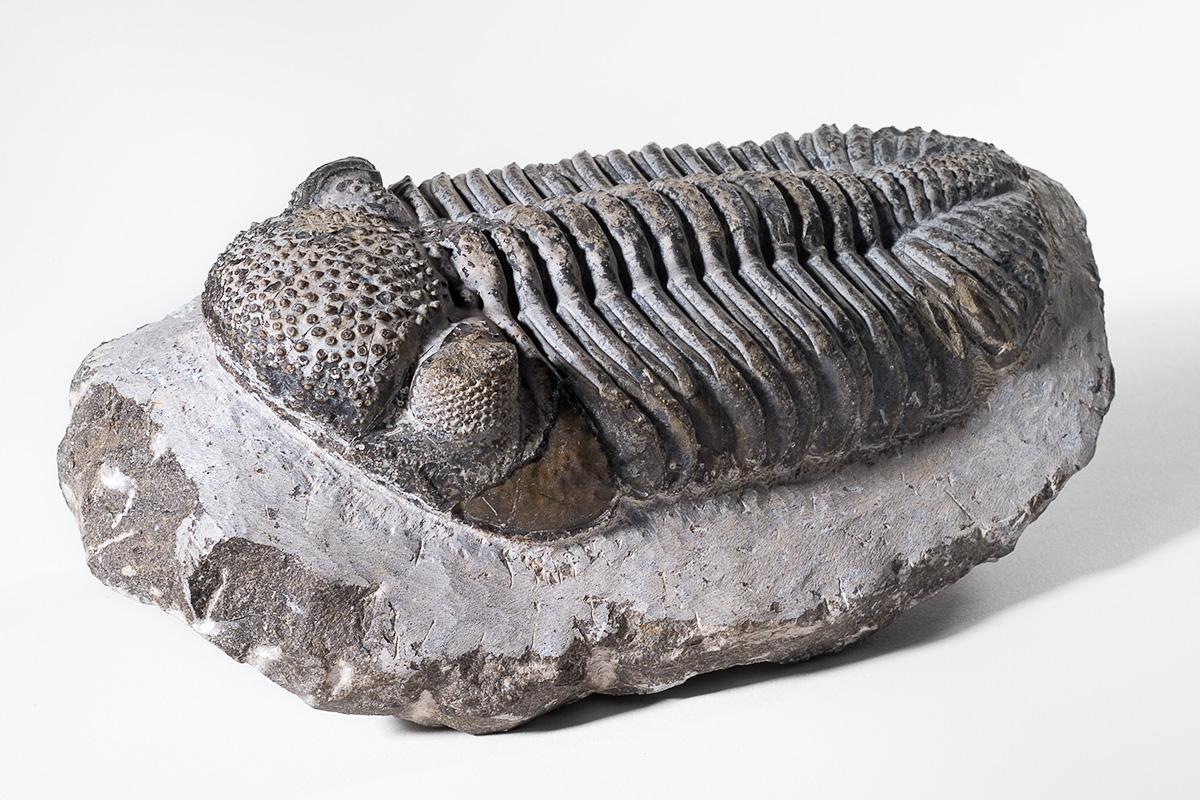
The eyes of trilobites were studied in a new paper published in the international journal Nature Communications by a team of scientists with Jason Dunlop from the Museum für Naturkunde. The authors used state-of-the-art techniques such as synchroton- and micro-CT on some exceptionally preserved fossils to reveal the internal anatomy of trilobite eyes. They confirmed that trilobites had structures called crystalline cones beneath the lenses of their multifaceted compound eyes. The fact that trilobites had these cones strongly suggests that they are indeed more closely related to myriapods, insects and crustaceans.
Trilobites are iconic fossils. These arthropods inhabited Palaeozoic oceans for more than 300 million years from the Cambrian through to the Permian, and more than 20,000 species have been described. Despite their abundance, and familiarity to fossil collectors, an unresolved question is the position of trilobites in the arthropod Tree of Life. On the one hand, they resemble horseshoe “crabs”, which are in turn related to arachnids, e.g. spiders and scorpions. On the other hand, trilobites had antennae which are also seen in insects, crustaceans, and myriapods. A further important feature which separates the living arthropods is the detailed structure of their eyes. An important question in arthropod evolution is whether trilobites are more closely related to a broad group called Chelicerata, i.e. horseshoe crabs and arachnids, or the Mandibulata which includes centipedes, millipedes, crustaceans and insects. The mandibulate arthropods have chewing mouthparts (i.e. mandibles), which are not present in trilobites, although trilobites and mandibulate arthropods do share the presence of sensory antennae. A further important difference between chelicerate and mandibulate arthropods is the way in which their eyes are constructed. All arthropods need to focus light from their eye lenses onto the light-sensitive cells beneath. Chelicerates do this by having a downwards-pointing, cone-like extension of the cuticle making up the eye lens, while mandibulates have evolved a special separate structure called a ‘crystalline cone’ made up of four translucent cells which again direct the light downwards. So which eye type do trilobites have? Scholtz et al. studied some exceptionally preserved trilobite fossils – Asaphus from the Ordovician and Archegonus warsteinensis form the Devonian – using techniques such as scanning electron microscopy and computed synchroton and micro-tomography. They could show convincingly that trilobites also have a crystalline cone, and even the light-perceiving region at the base of the cone called the rhabdom. The fact that the internal anatomy of these fossil arthropod eyes can be resolved is in itself an exciting result. It is all the more important for providing evidence that trilobites share a common ancestor with the mandibulate arthropods, all of whom evolved this more complex light-gathering system.
Free pictures here:
http://download.naturkundemuseum-berlin.de/presse/Trilobitenaugen
Image 1: Comparison of the eyes of living arthropods. Photo: Gerhard Scholtz.
Image 2: Cross section using scanning electron microscopy through the eye of the Devonian trilobite Archegonus warsteinenesis showing the lens (green), crystalline cone (blue) and rhabdom (purple); a structure similar to that of insects, crustaceans, centipedes and millipedes. Photo: Gerhard Scholtz.
