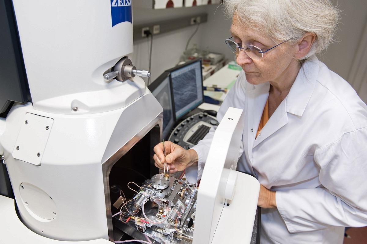
The museum's integrated zoological research laboratory facilitates gene expression and immunohistochemistry studies as well as histological and electron microscopical documentation of zoological objects. Spread over an area of about 700 square metres the laboratory provides ample space for large-scale equipment and the associated preparation labs. This helps to integrate different analytical methods and to optimise workflows. A close connection with the DNA laboratory is key in this respect. Section series can be prepared for light and electron microscopy, e.g. to study the evolution of certain organ systems or limb regeneration in vertebrates. Fluorescence and confocal laser scanning microscopes help visualizing cellular processes such as gene expression or protein distribution based on antibody- or RNA-based staining methods.
Methodology (selection):
- Histology
- Light and electron microscopy
- Immunohistochemistry
- Gene expression studies
Equipment (selection):
Scanning electron microscope ZEISS EVO LS 10
Technical Assistant: Anke Sänger
For the investigation of morphological surface structures. The microscope’s variable pressure function facilitates analysis of untreated samples. This is particularly important for examining collection material. The instrument is equipped with SE, VPSE and BSE detectors as well as a Pelletier element for sample cooling.
Transmission electron microscope LEO 906
Technical Assistant: Anke Sänger
Analysis of ultra-thin section series of tissues at subcellular level and subsequent 3D reconstruction of cell structures and organ systems. The microscope runs on 80 kilovolts and also covers a low magnification range (below 1K) without limitations. Images are recorded with a 2K CCD camera.
Confocal laser scanning microscope LEICA TCS SPE
To localize fluorescence signals in tissues. These signals either originate from the tissue itself (auto fluorescence) or from specific antibodies that are experimentally applied to detect certain proteins. The microscope is equipped with diode lasers exciting four different wavelengths including a UV channel.
Applications:
- The confocal laser scanning microscope was used to study the nervous system of brittle stars to test whether skeletal elements in the arms of the animals act as light conductors for underlying photoreceptors, as postulated in an article published in Nature in 2001. It was shown that photoreceptors are distributed over large parts of the animal's surface in three species of the genus Ophiocoma, but that no light-sensitive cells can be found in the supposed focus area of the skeletal elements described as microlenses. Irrespective of the physical light-conducting properties of these microlenses, they apparently have no function for the visual system of the animals.
- A multi-method approach using light and scanning electron microscopy (morphometric part) and molecular analysis (genetic part) provided evidence for hybridisation of the two Atlantic brachiopod species Terebratulina retusa and Terebratulina septentrionalis. This is remarkable, because the two species were apparently separated by the formation of the Atlantic and established as independent species about 60 million years ago. The region southwest of Iceland is influenced by the Gulf Stream and could be identified as the contact zone between the two species. The samples were collected during an expedition (Me 85/3 “IceAGE”) with the research vessel “Meteor”.
-
Furchheim (2016) Functional morphology of the light sense organs of recent Brachiopoda. Dissertation, Humboldt-Universität zu Berlin, 121 pp.
