This article was first published in our journal For Nature (issue 4/2021).
The latest computed tomography makes it possible: With the new CT scanner, researchers can see inside the animals – without destroying fine tissue
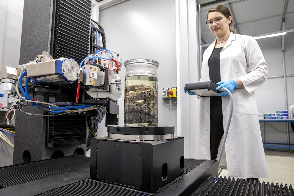
Kristin Mahlow, CT technician, enters the eight by eight meter "Kubix", a container with an unusual content: Here is the 30 tonne CT scanner FF85 from YXLON. It was specially made for the museum and is used to digitize the collections. This centerpiece of the system can scan objects up to 90 centimeters in size: antelope and gorilla skulls, which are digitized as part of the removal of the skull collection; Preparations soaked in alcohol, which can be examined non-destructively with skin and hair, or fossils. There is strong demand for CT from national and international partners, especially in times of pandemics, because it enables digital objects to be made accessible to research worldwide without researchers having to get on a train or plane. Mahlow will often go to the Kubix in the next few years, because the digitization of the collections is part of the museum's future plan.
Her favorites are snakes. She has been working on her doctoral thesis for two years. Bothrops jararaca is a Brazilian venomous snake, a pit viper that is responsible for almost 90 percent of bite accidents in cities on the east coast of Brazil. This species of snake changes food during its life: young animals eat amphibians, reptiles and insects; adult animals eat small mammals. The poison of the young is therefore composed differently than that of the adults. This is important for the development of antisera and was only recently discovered by researchers.

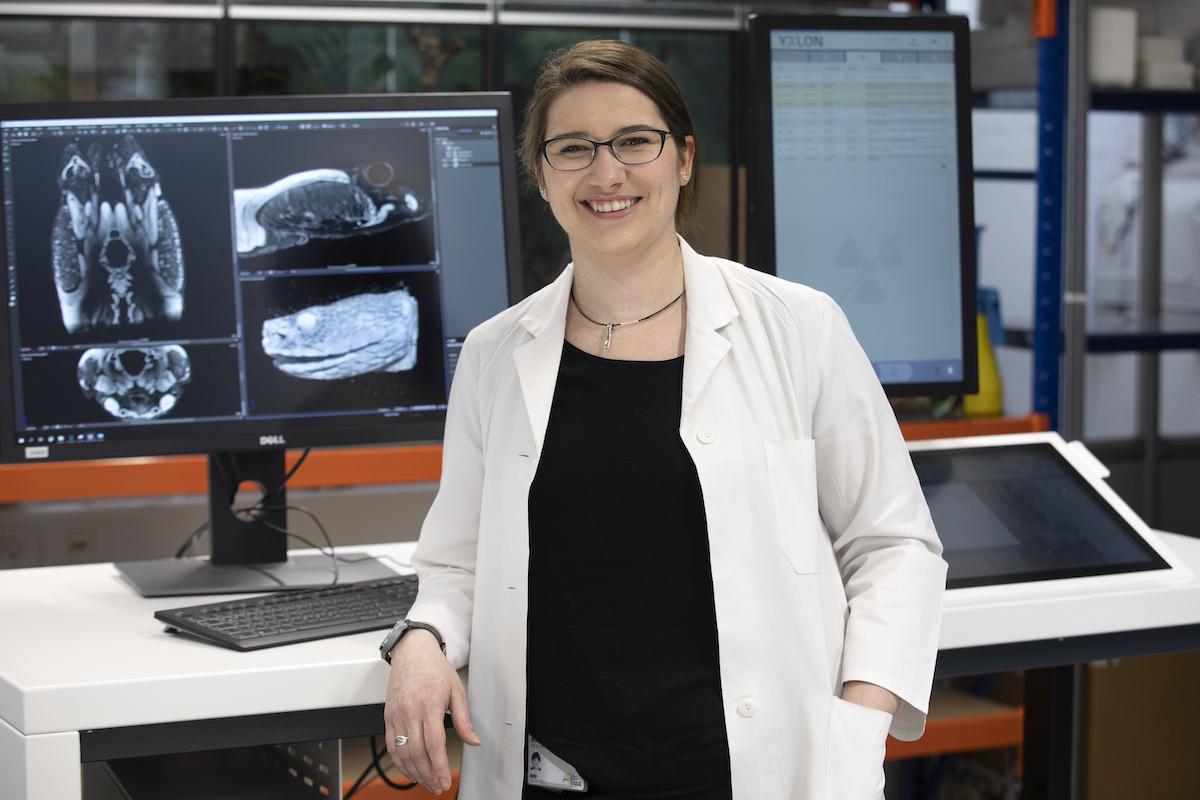
A research project in collaboration with the University of São Paulo deals with the morphological analysis of the venom glands: When the venom changes – what changes in the skull and in the venomous apparatus of the snake? Or does anything change at all? To research this, animals of all ages must be examined for their bone structure. To do this, Kristin first makes a standard scan. The specimens are then treated with an iodine staining solution, which makes the soft tissue of the head visible, such as the jaw muscles, the brain or the venom gland. The high-resolution CT scanner is required for these scans. A resolution far below the thickness of a human hair can be achieved.
And what do the snake scans show now? "The structure of the glands is different in young animals than in adult animals," says Kristin. "The gland cells change their relative size and position and structure themselves differently." How and why this happens is one of the research aspects of her doctoral thesis.
Text: Gesine Steiner
Pictures: Pablo Castagnola
